5 Spectral diagnostics of crop phytopathologies using trained models
The quality assessment of the trained classification model for crop phytopathologies is performed using new, previously unseen hyperspectral images of affected plant tissue regions. Multichannel spectral data enable detection of subtle spectral differences between healthy and diseased plant tissues by covering a wide range of wavelengths.
During the spectral diagnosis of phytopathologies, new hyperspectral images are analysed using a classification database built from previously identified infected regions, according to the trained model designed to detect new infections (Figure 88). To achieve this, it is initially necessary to create a separate folder for importing the new images by clicking the “Add” button in the lower-left corner of the panel and entering the name of the new folder as “Test” (Figure 89).
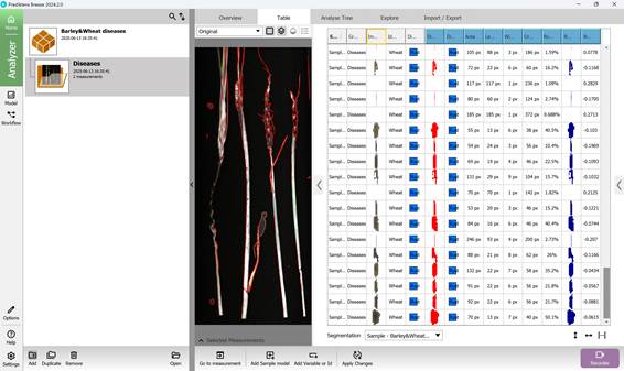
Figure 88 – Classification database of infected crop regions created using the trained model for detecting new infections
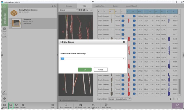
Figure 89 – Creating a test folder for importing newly acquired hyperspectral images
The import stage also includes preprocessing within the Image import wizard, consisting of calibration, noise removal, and image segmentation steps (Figures 90, 91). Segmentation is performed based on the previously created reference (baseline) model. Then, a classification algorithm based on the PLS-DA model is applied. Using spectral features previously associated with specific diseases, the algorithm identifies the type of infection if present. This enables obtaining the results of applying the classification model to new hyperspectral images and diagnosing crop infections caused by “Loose smut” and “Brown rust” pathogens (Figures 92, 93). Such differences are often undetectable using standard visual methods or visible spectrum cameras.
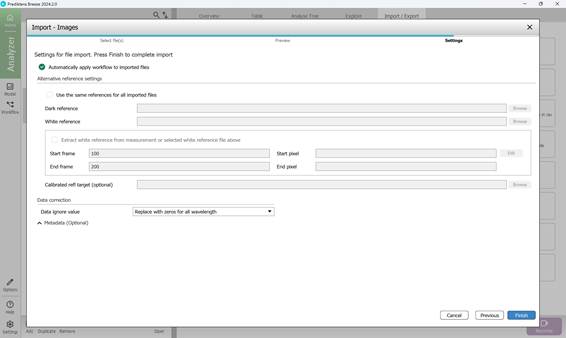
Figure 90 – Importing new hyperspectral images for preprocessing and calibration
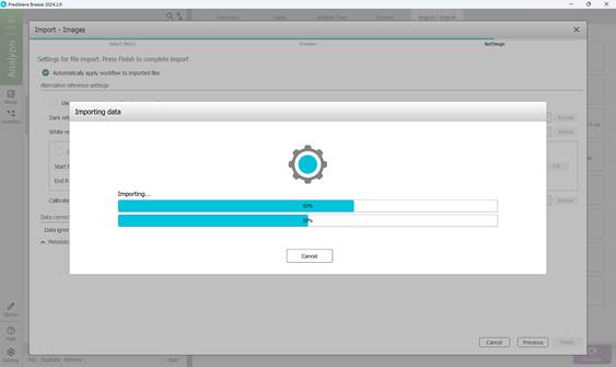
Figure 91 – Preprocessing and calibration of new hyperspectral images featuring crop disease samples
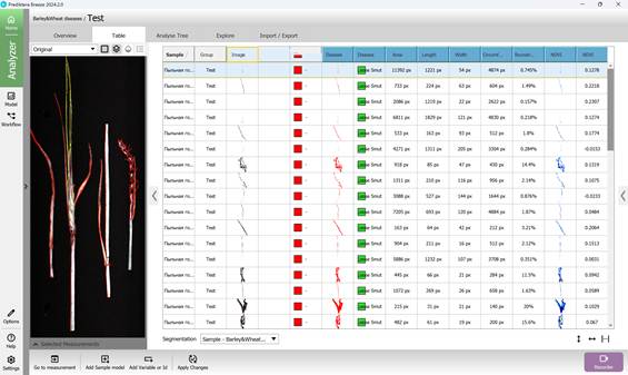
Figure 92 – Results of applying the classification model to new hyperspectral images for diagnosing damage caused by “Loose smut” pathogens
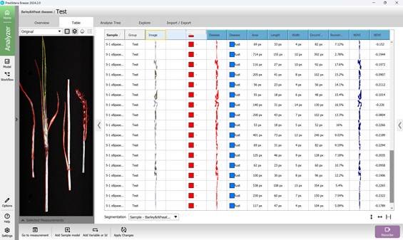
Figure 93 – Results of applying the classification model to new hyperspectral images for diagnosing damage caused by “Brown rust” pathogens