5 Physiology of sensory systems
All variety of phenomena and objects of the objectively existing world is perceived by special senses - analyzers, which consist of three parts: peripheral, conductive nd cortical. The peripheral part is represented by specific receptors (ear, nose, eyes, skin, etc.) that perceive certain types of stimuli; the conductive part transmits impulses to the central nervous system; processes of analysis and synthesis of stimuli coming from outside are carried out in the cortical part. The visual and dermal analyzers are a powerful source of information. With the help of the visual analyzer light and color sensations are perceived, whereas skin analyzers give the opportunity to feel tactile, warm, cold, and painful irritations.
5.1 Physiology of analyzers
The purpose of the lesson: Identify the presence of a blind spot on the retina of the eye and the field of vision; study the tactile sensitivity of the skin and to determine the spatial threshold of this sensitivity; determine the sensitivity of individual parts of the tongue to various taste irritations.
The following is necessary for work: Mariott's drawing, perimeter of Fester, Frey hair, quinine solution, sugar, table salt, glass sticks, citric acid, a pair of compasses, rulers, chemical pencils.
Work 1 Experiment showing the presence of a blind spot on the retina
Work progress: At a distance of 20-25 cm from the eye Mariott's drawing is placed. The right eye is closed with a hand and the first image is traced with the left eye. By pushing the drawing away or drawing it closer, one can notice that at some distance from the eye the left image disappears due to the fact that its image falls on a blind spot (Figure 5.1
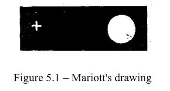
The experiment is repeated, closing the left eye and looking at the image with the right eye; in this case the right image disappears. Mariott's experiment is repeated.
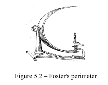
Work 2. Visual field
The visual field is a space within which all its points are visible at a fixed position of the eye. For the rays of different wavelengths, the field of view is not the same. The visual field is greatest for white color, i.e. for mixed light. This is due to the fact that the rods, sensitive to all visible rays and that perceive not the color, but the light, are also on the extreme periphery of the retina, where there are no cones - the apparatus of color vision. To determine the visual field, the perimeter of semicircle is used, the perimeter is graduated in degrees. A special plate serves as a support for the chin of the subject. In the middle of the semicircle of the perimeter there is a mirror, which the subject fixes with their eye.
Work progress: To start the experiment, draw a diagram in the notebook, shown in the figure. Ask the subject to put their chin on the plate of the perimeter (Figure 5.2), close one eye, and to trace the mirror the other. Follow the perimeter scale from the periphery to the center, first from the top down, and then from the bottom up, the slider in with a colored circle. Note on which degree the subject began to clearly see the color offered for distinguishing. The experiment is carried out first with the vertical position of the semicircle, and then when it is rotatedto 45, 90, 135, 180 degrees. Test colors: green, red, blue, and white. The subject should not know in advance what the color of the slider on the scale. Therefore, the colors in the experiment must be changed all the time. Mark on the diagram drawn in the notebook the distances from the center in degrees, on which the subject was able to distinguish a particular color. Connect the dots of each color to get the curves that limit the visual field for the studied colors. Repeat the same for the other eye.
Work 3. Detection of the thermal, cold and tender points of the skin
The frequency of location of thermal, cold and tender points on the same surface area of the body is not the same. On average, 1 сm2 of the skin surface has 50 tender, 25 tactile, 12 cold and 1-2 thermal points.
Work progress: (Work is carried out by two people). Find thermal and cold points on the back surface of the wrist and radiocarpal joint using heated and cooled pinheads, and mark them with multicolored ink (Figure 5.3).
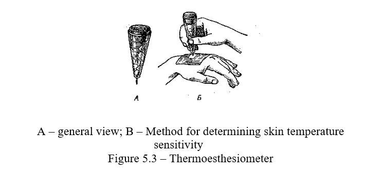
Then, with the tip of the pin, find and mark the tender points. Calculate and compare the frequency of location of thermal, cold and tender points on 1 cm2 of skin surface.
Work 4 Determination of the sensitivity of individual parts of the tongue to various taste irritations
The taste receptors of the tongue perceive bitter, sweet, sour and salty tastes. Different parts of the tongue have a different ability to perceive these taste irritations. Thus, the tip of the tongue is most sensitive to sweetness, its edges to acidic, the root to bitter, the tip and edges to salty, the middle part of the tongue's back is very low in sensitivity to all taste irritations.
Work progress: On different parts of the subject's tongue, apply a drop of quinine, sugar, table salt and citric acid solution with a glass rod. The subject should not know in advance what solution is applied to one or another section of the tongue, their task is to determine the taste of the solution. During the break between individual determinations, which must be at least 2 minutes, the subject rinses the mouth well with distilled water. Based on the answers, draw a map of the taste buds of the tongue (Figure 5.4).
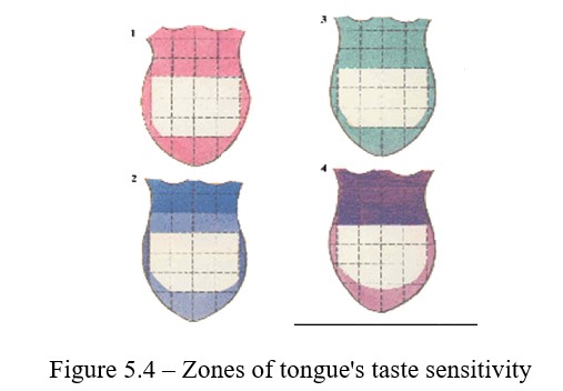
Identify and sketch areas of taste sensitivity of the tongue.
Work 5 Determination of Acuity of Hearing
Purpose: Using a wrist mechanical watch to determine the severity of the hearing.
Equipment: mechanical wristwatch or stopwatch. The sensitivity of the ear to sound stimuli is called acuity of hearing. In healthy people, it can be different. Examine the acuity of hearing with the help of quiet and loud speech or with a mechanical clock, tuning fork. The threshold of hearing is the minimum loudness that can be perceived by the ear of the subject. Hearing is measured in decibels. Auditory sensitivity is the inverse of hearing threshold. A hearing is considered normal, when the sensitivity threshold is 10-15 cm.
Work progress: 1. Determine the severity of hearing. Put the mechanical watch to your ear and push it away from you until the ticking disappears. When the sound disappears, measure the distance between the clock and ear. This experience proves auditory sensitivity, ie, the analyzer is able to adapt to the stimulus. The greater the distance between the clock and ear, the better the auditory sensitivity. Now move the arm clock to the arm's length and slowly bring them closer to your ear until a barely noticeable sound. Measure this distance - this is the threshold of hearing. Calculate the average figure between these indicators: the threshold of hearing and auditory sensitivity.
2.Recommendations on execution of the protocol of work
The obtained results of the study should be written down in the notebook of the experimental reports, compare them with the average indicators for the group.
Control questions
- The main functions of the analyzers and their classification?
- Olfactory apparatus?
- Gustatory analyzer?
- Cutaneous analyzer?
- Muscle and joint receptors (proprioceptors)?
- Auditory analyzer?
- Vestibular analyzer?