8 Physiology of the cardiovascular system
The heart provides a blood flow along the vessels. Its pumping function is based on the alternation of contraction (systole) and relaxation (diastole) of the heart. A human heart contracts about 70 times per minute. By determining such indicators of blood circulation as the rhythm of heartbeats, the minute volume of the heart, the level of blood pressure, etc. one can judge the physiological state of a human and animal, the organism's adaptation to certain changes in the external and internal environment. The heart of a frog consists of three parts: two atria and one ventricle. Each cycle of cardiac activity of a frog is composed of four phases:
1) systoles or contractions of venous sinus;
2) atrial systoles;
3) ventricle systoles and its diastoles;
4) general pause or est of a heart.
There is no venous sinus in the heart of warm-blooded animals; therefore, three phases are distinguished in the work of the heart: the systole of the atria and ventricles, the diastole of these sections and the general pause. A simple observation of the work of the heart does not provide a fine analysis of their activity, so the method of graphical registration is used instead. The obtained cardiogram allows to study many aspects of the heart activity: the frequency and strength of contraction, the sequence and duration of cardiac contractions.
8.1 Recording of frog's cardiac activity and cardiogram analysis
The purpose of the lesson: Visually determine the sequence of contractions of the cardiac divisions, record a cardiogram, and analyze it.
The following is necessary for work: frog, kymograph, universal tripod, needles, preparatory boards, preparation set, double heart lever, two serfins, Ringer's solution for cold-blooded.
Work 1 Recording frog's cardiac activity
Work progress: Cut off the upper jaw of the frog with the brain and destroy the spinal cord. Then fix the immobilized frog (abdomen facing upward) with needles on the dissection board. Open the thorax: cut the skin 1-2 cm away from the pelvis along the midline of the abdomen to the lower jaw, the second transverse incision is made at the level of the shoulder girdle, and the formed triangular rags of the skin should be removed. Then grasp the sternum with forceps, lift it, cut the surrounding tissues of clavicla and remove them. The contracting heart is clearly visible. The heart sac is pulled off with forceps and cut with scissors. The bridle is cut. If the heart is raised with forceps, a venous sinus that contracts frist, is clearly visible (Figure 8.1)
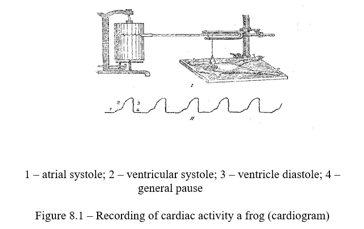
The whole cycle of the heart is traced. Then a graphical recording of the heart starts: the dissecting board is fixed in a tripod under the Engelman's lever; the top of the heart is grasped with a serphin, connected by a string with a short arm of Engelmann's level. The contraction of the heart leads to the lifting of the lever, and relaxation leads to the lever dropping. Movement of the lever is recorded on the surface of the kymograph. Small contractions, atrial systoles can be observed on the kymograph, and then large ones - ventricular systoles. The resulting kymogram is sketched and analyzed.
Control questions
- Basic parts of the circulatory system?
- What is the dependence of the force of the heart contractions on the stretching of the heart muscle fibers?
- Pumping function of the heart. Change in volume and pressure of blood in the heart cavities in different phases of the cardiac cycle?
- What are the ways to determine the minute and systolic volume?
- Cardiac valve and its meaning.
8.2 Heart automatism and its dependence on temperature
The ability for automatism is due to the presence of atypical muscle tissue in the heart. It creates and conducts excitation from one part of the heart to another. This tissue is called the conductive system of the heart. Clusters of this tissue form a sinus node in the venous sinus, and form a bundle of His in the atrioventricular septum. The leading part of the heart, which determines the heart's basic rhythm, is the sinus node. When it is turned off, the heart begins to contract due to impulses of excitations arising in the underlying sections of the conducting system (Figure 8.2).
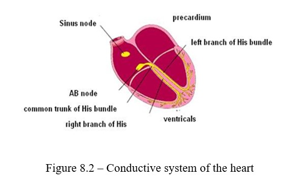
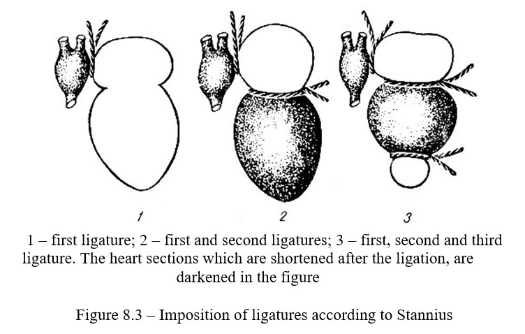
Conductive system of frog's heart consists of 3 nodes: Remak, Bidder, and Ludwig. This is especially evident in the heart carved from the body. The leading role of the sinus node (the Remak node) is observed in Gaskell's experiment and by imposing the Stannius ligatures (Figure 8.3).
The purpose of the lesson: to make sure that the heart muscle has automatism (as one of the properties of the heart muscle) and that the heart automatism is connected with the conduction system of the heart and there is a "decreasing" gradient of the automatism in the heart.
The following is necessary for work: frog, kymograph, universal tripod, needles, preparatory boards, preparation set, serphins, saline solution
Work 1 Analysis of the conductive system of the heart (the Stannius experiment)
Work progress: The frog is immobilized and the heart is exposed in the usual way. The thread is put under the heart using forceps and the bridle is bandaged, which is then cut below the knot. The top of the heart is grasped with a serphin connected to Engelman's lever and the frequency of heart contractions is counted. The first Stannius ligature is applied to the heart between the venous sinus and the atrium, for this, a 13 to 15 cm thread is under the aortic branch. At first, the ligature is tightened slowly, then quickly, strictly on along the white strip which delimites the sine and atria. The significance of the individual parts of the conducting system can be studied by imposing ligatures (filaments) on the heart of the frog according to Stannius. The heart stops immediately, and the sine continues to contract. The number of contractions of the venous sinus is counted. The second ligature is applied between the ventricle and the atrium; this causes the restoration of contraction of either the ventricle, or the atria, or both, together, depending on the location of the ligature with respect to the Bidder node.
Work 2 The influence of temperature factors on the work of the heart
Work progress: The frog is immobilized and the heart is opened in the usual way. The top of the heart is grasped by a serphin, which is connected to the Engelman's lever. The cardiac contractions per minute are counted; then a thermode with cold water is applied to the venous sinus and heart contractions heart rate per minute (each) is counted for 2-3 minutes. After the restoration of the number of heart rate, an experiment is carried out with the application of a thermode with warm water to the venous sinus.
Control questions
- What options in reducing the individual parts of the heart are possible with the application of the second ligation of Stannius?
- What is the speed of propagation of excitation in the working and conductive musculature of the heart?
- What is cardiac blockade and fibrillation??
- Automatism of the heart. The gradient of automatism (the Stannius experiment). Modern ideas about the substrate and nature of automatism?
- Modern ideas about the substrate and nature of automatism.
8.3 Extrasystoles and cardiac biocurrents
In the heart muscle as well as in any other muscle, after each outburst of excitation, there is a period of complete inexcitability (absolute refractivity). During the whole period of contraction, the heart does not respond to any stimuli. When there is relaxation of the cardiac muscle, the excitability of the heart muscle appears and gradually increases. However, in the initial period of relaxation, excitation is low, which gives reason to call this state as a relative refractivity. In the stage of relative refractivity, excitation impulses do not usually occur in the conducting system. But if you apply a strong stimulus (for example, needle pricking) to the area of the sinus node, excitation appears in the cardiac muscle and the heart contracts. An extraordinary contraction of the heart is called an extrasystole. After the extrasystole there is an extended pause, called a compensatory pause. The excitability of the heart muscle during the diastole of the heart sharply rises (the phase of exaltation). Extrasystole is a non-regular systole that occurs in response to irritation at the time of the relative refractivity phase. Normally, it does not ossur, but it can be obtained in an experiment (Figure 8.5).
The purpose of the lesson: investigate the excitation of the heart muscle in various phases of its activity. Prove and study the mechanism of occurrence of electrical phenomena in the heart muscle
The following is necessary for work: Frog, kymograph, universal tripod, Murrey's cardiographic forceps, preparation set, serphins, physiological solution, electrostimulator, Ringer's solution, ligatures.
Work 1 Observation of the biocurrents of the heart (the Kelliker’s experiment)
One of the indicators of the heart work is its biocurrents. The biocurrents of the heart can be detected biologically. One such method is the Kelliker's experiment.
Work progress: The chest of the rat is opened and the heart is uncovered. The neuromuscular medication is placed on a slide, and its sciatic nerve is put onto the contracting heart using a glass hook. At this time, the gastrocnemius muscle contracts in one rhythm with the heart muscle, but the leg contraction occurs earlier, which is explained by the fact that the duration of the latent period of the skeletal muscle (1.01 sec) is shorter than that of the cardiac muscle (0.5 sec). It is necessary to record the results, sketch out the scheme of the experiment and draw conclusions.
Work 2 Dynamics of changes in the excitability of the heart muscle and the extraction of extrasystoles
Work progress: An apparatus for applying single stimuli to the heart is assembled. The frog is immobilized and fixed on a plank, and its heart is opened in the usual way. The plank with the frog is fixed in the tripod. The ventricle of the heart is placed in the cardiographic forceps. The recorder of the cardiographic forceps is tangentially approximated to the surface of the kymograph and the curve of the normal heart contraction is recorded (Figure 8.4).
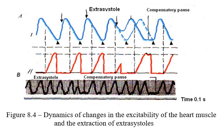
The threshold of muscle irritation is determined with the help of the electrostimulator. The strength of the current is increased by approaching the coils and single stimuli are applied in a sign of the initial cardiogram at various periods of cardiac activity: at the beginning of the systole, in the middle of the systole, at the beginning of the diastole, during a general pause.
Control questions
- What is absolute and relative reactivity?
- How does the excitability of the cardiac muscle change during the cardiac cycle?
- What is the duration of the absolute refractory period for the muscles of the atria and ventricles?
- What are extrasystole and compensatory pause, what are the causes of their occurrence?
- When do biotics appear in the heart and how can they be detected?
8.4 Electrocardiography
Electrocardiography is an objective method for studying cardiac activity. Electrocardiography, as a method, is widely used to understand the nature of the onset and spread of excitation and determination of the causes of disorders in the heart activity. The method is based on a graphic record of the electric currents that arise when the heart is excited, the force lines of which propagate throughout the body. For recording biocurrents a device called electrocardiograph is used; its electrodes are applied to certain parts of the animal's body.
The purpose of the lesson: озакомиться с методом регистрации биоэлектрических явлений в сердце (электрокардиографией).
The following is necessary for work: electrocardiograph, subject, 10 percent sodium chloride solution, gauze wipes
Work 1. Electrocardiography (ECG)
Electrocardiography, i.e. the registration of bioelectric phenomena accompanying the activity of the heart, allows one to examine cardiac activity and to recognize its violation, which is of great importance both for studying the work of the heart in various conditions (especially in physiology of sports) and for medical practice. Successive changes in the potential difference between individual parts of the heart muscle, associated with the course of the cardiac cycle, can be recorded with a special oscilloscope (electrocardiograph). The body is a good conductor. Therefore, the shifts in the potentials of the working heart can be detected if the outgoing electrodes are applied not only directly to the heart, but also to the surface of the body. This makes it possible, without complex procedures and without causing painful sensations, to record an electrocardiogram of a human. The skin areas, to which the lead electrodes are applied, are different. There are so-called leads (at least one of these electrodes is applied in the region of the location of the heart). The most widely distributed leads from the limbs are the following three (Figure 8.5): I - from the left and right hands; II- from the right hand and left leg; III - from left hand and left leg.

Excitation spreads through the heart in various and changing directions, and therefore, the electrocardiograms obtained with different leads are not the same. Each cardiac cycle of the electrocardiogram has five main teeth. With the usual recording, the upwardly directed deflections show the electronegativity of the base of the heart, and the downward directed ones show electronegativity of the apex of the heart. The deflections are denoted by Latin letters. The deflection reflects the systole of the atria, and the QRST-systole reflects the systole of the ventricles and is called the "ventricular complex."
The following is necessary for work: an electrocardiograph, a couch.
Work progress: For the study, use an electrocardiograph of any brand or an oscilloscope. Recently, the ELKAR-3 ink-recording electrocardiograph, which connects to a power supply, is widely used in the clinics. Before applying the electrodes to the areas, wipe them with alcohol. Tape the electrode firmly. Turn on the electrocardiograph, record the electrocardiogram. During the recording, the subject must lie quietly without straining their muscles. Develop the film, sketch the electrocardiogram in the notebook and analyze the heart cycle.
Control questions
- What is the method of electrocardiography based on?
- The nature of the ECG and the origin of its deflection?
- ECG leads. Human ECG analysis in norm (the shape and size of the ECG deflection, the duration of the intervals, the electric axis of the heart)?
- The cycle of the heart work and its phases. Phase analysis of the cardiac cycle. Cardiac valve and its significance?
- Pumping function of the heart. Change in volume and pressure of blood in the heart cavities in different phases of the cardiac cycle?
- Heart tones and their origin. Phonocardiography?
- Vascular system. The basic laws of hemodynamics?
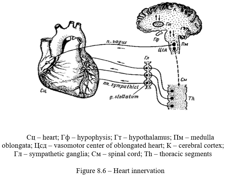
8.5 Nervous regulation of cardiac activity
The work of the heart is regulated by the autonomic nervous system (vagus and sympathetic nerves) (Figure 8.6), with the stimulation of which it is possible to observe 5 different influences:
1) Changing of the heart rate (chronotropic);
2) Change in cardiac contraction force (inotropic);
3) Change in the tonus of the heart muscle (tonotropic);
4) Change in excitability (bathmotropic);
5) Change in conduction of the heart (dromotropic).
Similar results are obtained by stimulation of the nerve centers of these nerves.
The purpose of the lesson: determine the influence of sympathetic and vagus nerves on the work of the heart.
The following is necessary for work: frogs, board for fixing a frog, preparation set, ligatures, electrostimulator, salt crystals.
Work 1 Irritation of the vagus nerves center
Work progress: Frog, without immobilization, is wrapped in a cloth and the brain is exposed, picking out the cartilage (behind the eyeballs) to the top side using scissors and forceps. Find the rhomboid fossa and cover it with a cotton tampon soaked in Ringer's solution. Then attach the frog to the dissection board with its abdomen up, expose the heart and count the number of contractions per minute. After that, lay a crystal of table salt in the medulla oblongata region and again count the number of cardiac contractions every 2 minutes until the original rhythm is restored. After the experiment, write down the results and draw conclusions.
Work 2 Irritation of the vagosympathetic nerve of a frog
Work progress: The thoracic cavity of the immobilized frog is opened and the heart is widely exposed. The pericardium is removed and the bridle is cut. Firstly, the right forelimb is pulled to the side and down. The muscles and fascia are cut between the angle of the jaw and the heart using scissors. This way gives access to the armpit. Using a small cotton tampon at the tip of forceps and a preparation needle, a vascular-neural bundle, consisting of a cutaneus-pulmonary artery, glossal, glossopharyngeal, laryngeal, and vagosympathetic nerves, is found. Two nerve trunks distinctly protrude and lie ahead. The most anterior-glossal, the second-glossopharyngeal, which makes a loop behind these nerves, pass the vagus and guttural nerves. The vagus nerve is situated more deeply and is the most posterior.
The vagosympathetic trunk is released from the surrounding tissues (the rest of the nerves are cut) and thread is put under the nerve. An electrical circuit is made, the battery is connected to the induction coil via a breaker, and electrodes for nerve irritation are attached to the terminals of the secondary coil. The heart rhythm is counted, the peripheral segment of the vagosympathetic trunk is gently lifted by the thread, the electrodes are placed under the segment and irritation begins, closing the key of the primary circuit. The rhythm of cardiac contractions is counted when the vagus nerve is irritated, and also after the removal of the stimulus.
Work 3. The influence of the sympathetic nerve on the heart work
Work progress: To find the sympathetic nerve, look for the thoracic aorta (left or right), located on the sides of the spine. A thin, gray sympathetic chain is closely adjacent to them. This chain is taken for ligature near the celiac vessel and the caudal node is cut. The preparation is done under the magnifier to the point where the sympathetic chain crosses the brachial plexus. This segment of the sympathetic nerve is placed on thin electrodes. The frequency of contraction of the heart is counted before and at the moment of irritation of the sympathetic nerve. After the experiment, record the results and make conclusions.
Control questions
- Which nervous system regulates the heart function?
- Where are the centers of cardiac innervation?
- How does the work of the heart and the properties of the heart muscle change during irritation of the sympathetic and vagus nerves?
8.6 Reflexes of the heart
The most diverse external and internal stimuli cause shifts in the work of the heart. Influence from extera- or interoreceptors on the activity of the heart is carried out reflexively, by changing the tonus of the vegetative centers. Reflex effects on the heart from the internal organs can be observed in the experiments of Golts, Ashner-Danini and from the blood vessels in Goering's experiment.
The purpose of the lesson: to be convinced in the presence of reflex influences on cardiac activity and blood pressure
The following is necessary for work: frogs, preparation set, fixing board, cotton wool.
Work 1. Golts’ experiment
Work progress: The frog's brain is removed, except for the oblong one. To do this, cut off the upper jaw behind the eyes. Fix the frog, with its abdomen up, on the plate, cut a small patch of sternum over the heart and expose the heartStop the bleeding with cotton tampons. Count the number of cardiac contractions in 1 minute. Then lightly tap on the abdominal wall with forceps and count the heart rate. After the normalization of the work of the heart, destroy the spinal cord and tap on the abdominal wall again and observe the heart rate. In the end of the experiment, record the results and draw conclusions about the place of closure of the reflex arc.
Work 2 Ashner-Danini reflex
Work progress: Check the subject's pulse rate on the radial artery per minute. Apply both hands to the side surface of the subject's head, and ask them to close their eyes, slowly presses both eyeballs simultaneously for 5-8 seconds with your thumbs (not too much) and quickly stop pressing. Check the pulse rate and compare it with the original number. Write down the result of the experiment and draw a conclusion about the reflex arc of the Ashner-Danini reflex.
Control questions
- What do you know about the reflex activity of the heart?
- Is it possible to conduct the Golts' experiment on a frog with a destroyed spinal cord?
- How can we prove that the inhibition of the rhythm of contractions and even the arrest of cardiac activity observed during irritation of the peritoneum is due to an increase in the tonus of the vagus nerve?
- What is the path of the reflex arc of the Golts' reflex?
- How do we explain the mechanism of changes of the heart work and blood pressure in the Goering's experiment?
8.7 Humoral regulation of cardiac activity
The influence of hormones, electrolytes and mediators on the activity of an isolated heart. The activity of the heart can change under the influence of various chemicals: hormones, salts and other humoral factors carried by blood (Figure 8.7). To maintain the normal activity of an isolated heart, the Ringer's solution is used. The decrease or increase, as well as the change in the ratio of the ionic composition in these solutions change the cardiac activity.
The purpose of the lesson: to study the effect of electrolytes - K and Ca of some hormones and mediators on the work of the heart
The following is necessary for work: frogs, kymograph, Engelman's levers, serphins, preparatory set, ligatures, Straube's canula, K and Ca solutions, pipettes, Ringer's solution, adrenaline solutions and acetylcholine.
Work 1 The influence of hormones, electrolytes and mediators on the activity of an isolated heart
Work progress: The frog is immobilized on the dissection board, the heart is exposed in the usual way. The heart is then isolated by the Straub method. For this, ligatures are put under both arteries and under the aortic bulb. The first two ligatures are tied. An oblique incision of the left artery is made and the Shtaraube cannula, previously soaked in Ringrenra's solution, is injected. The cannula is injected into the ventricle, without damaging the spiral valve. Then the heart is carefully cut out from the body, trying not to damage the venous sinus. The blood is pumped out from the heart with a bulb syringe; the heart is rinsed with Ringer's solution to avoid formation of clots. An isolated heart is fixed in a tripod, and the top of the heart is grasped by a serphin and connected to the Engelmann's lever. The work of the heart is recorded on the surface of the kymograph under the action of various solutions in the following sequence:
- ) The kymogram is recorded and the heart rate of an isolated heart is counted during nourishment with Ringer's solution;
- ) Ringer's solution is sucked out with a bulb syringe and the canula is filled with Ringer's solution with an excess of Ca ions, a kymogram is recorded, and the heart rate is counted;
- ) Ringer's solution with excess Ca is sucked out with a bulb syringe. The heart is rinsed with the usual Ringer's solution and the heart is restored using Ringer's solution with an excess of K ions. A kymogram is recorded, and contractions of the heart are counted;
- ) Ringer's solution with excess K ions is sucked out; the heart is rinsed with Ringer's solution, and the heart's initial level of work is restored.Then the heart is filled with Ringer's solution with the addition of a few drops of epinephrine (1: 1000). A kymogram is recorded, and contractions of the heart are counted (Figure 8.7).
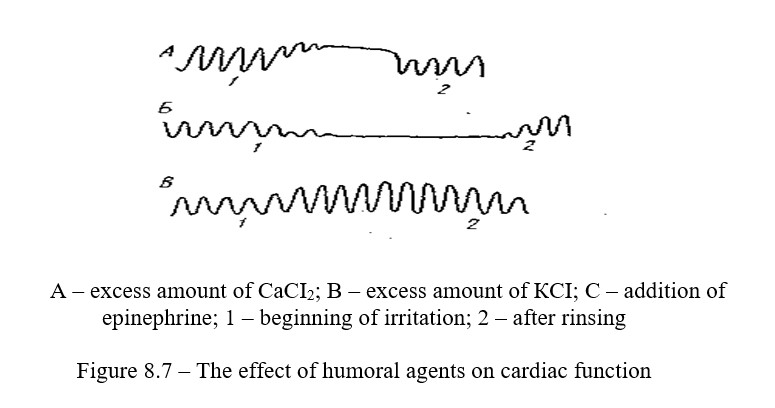
5) The solution with epinephrine is sucked out and the cannula is rinsed with the usual Ringer's solution; the original heart contractions are restored, and the cannula is filled with Ringer's solution with the addition of a few drops of acetylcholine. A kymogram is recorded, and contractions of the heart are counted (Figure 8.7).
Record the received digital data and sketch a kymogram. Make a conclusion.
Control questions
- What is humoral regulation of the heart?
- What are mediators, where do they stand out and what effect do they have on the work of the heart?
- How is the lumen of blood vessels regulated?
- What nerves are involved in the regulation of the lumen of blood vessels?
- What substances extend and contract the blood vessels?
- Name the humoral effects on the lumen of the vessels?
- How do K and Ca ions affect heart function?
8.8 Capillaroscopy and arterial pulse recording
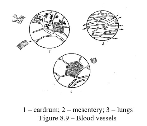
The nature of the blood flow in various parts of the vascular system is different. Blood is discharged in portions from the heart into the aorta, but then it moves along the aorta and arteries with a continuous current. Then the speed of its movement slows down. So if the blood flow velocity in the aorta reaches 500-600 mm/sec, in the arteries it decreases to 150-200 mm/sec, and in the arteriolas it drops to 5 mm/sec. In arteries and arteriolas, the blood flow is divided into two layers: the central (axial), where mainly the erythrocytes move, and the peripheral (plasma), formed by the blood plasma and leukocytes. The blood flow in the arteriolas is uneven, which is due to pressure pulse fluctuations. In the capillaries, the blood velocity decreases to 0.5 mm/sec, which creates favorable conditions for carrying out metabolic processes between blood and tissues.
In venules and veins, the blood flow velocity increases to 60-140 mm/sec, and in hollow veins it increases up to 200 mm/sec. The blood flow in the venules is uniform, without division into the axial and plasmatic layers. When observing blood circulation under a microscope, arterioles, capillaries and venules can be distinguished by the speed of blood flow in them, by the nature of the blood movement, and also by the direction of blood movement. In the arteriolas, the blood moves toward the branching, and is divided into axial and plasma layers, and its current is unevenly pulsating. In venules, the blood moves in the direction opposite the branching to a larger vessel, its current is uneven and continuous, without division into layers. The capillary walls are usually not visible, and chains of erythrocytes, moving one after another, can be seen.
The purpose of the lesson: Observe the peculiarities of the blood movement in the arteriolas, capillaries and veins of the live frog, under a microscope.
The following is necessary for work: frog, plank with holes, scissors, anatomical forceps, physiological solution, pipette, 0.5 liter can, 10% alcohol solution, cotton wool, microscope, tripod with clamp
Work 2 Capillaroscopy
Work progress: The frog is anesthetized by immersion in 10 % alcohol solution for 10 minutes. Then it is fixed on a plank. The paddle of one of the hind legs or tongue is stretched under the hole in the plank (Figure 8.8). The plank is placed on the microscopic stage so that the hole with the paddle or tongue is under the lens. The following observations are made:
1) Under a small magnification, arteries, capillaries and veins are found and drawn. Under the microscope, the arteries differ from the veins in that the arteries branch out along the course of the blood flow, and the veins aggregate. Capillaries differ in caliber, their diameter is equal to the diameter of erythrocytes, so the erythrocytes move in chain; during the passage of the bends of the capillaries, the erythrocytes are deformed;
2) Note the difference in the movement of blood in different parts of the circulatory system (speed, regularity);
3) Note the different speeds and axial parietal blood flow;
4) Inject 1 ml of epinephrine (0.01 percent solution) subcutaneously in the back and watch the blood flow rate;
5) Irritate sciatic nerve by induction current for 10-15 seconds.
At the end you need to sketch the arteries, capillaries, veins, write the results and make conclusions.
Work 3 Record arterial pulse (sphygmography)
Sphygmography - a method that allows analyzing the work of the left heart and the condition of the vascular wall. Record of arterial pulse can be done with the help of sphygmoplethysmograph with a hydraulic transmission consisting of receptive capsules, registering parts and the connecting tube with outgrowth to create an optimal pressure in system.
Work progress: hand of a testee need to be placed on a wooden stand of sphygmoplethysmograph. Fix the wrist with the strap and press slightly the sensor to the pulsing place with the help of screw with a curved metal plate at the end. With the help of a pressure pump creates the optimal pressure in the system at which the rubber membrane manometer begins to oscillate. Then the transmission system disconnects by the tap from pressure pump and record sphygmomanometry on a moving surface of kymograph.
Recording of the sphygmogram at a relative rest, after working with a free hand at the time of holding the breath on inspire and expire during hyperventilation of the lungs. Conduct an analysis of the sphygmogram: determine the rhythm of the pulse, the number of pulse waves per minute, the height of the pulse waves, the magnitude of the slope of the anacrotic limb and decaying limb.
Control questions
- What is the rate of blood flow in the arteries, veins and capillaries?
- What is meant by the linear and volumetric blood flow rate?
- What is the pulse, the types of pulse? Decaying limb? Anacrotic limb?
- What is the nature of the dicrotic wave?
- Explain continuity of a blood flow?
- Give a description of the blood flow in the capillaries?
- How can arteries and veins be identified in organs accessible to observation?
8.9 Determination of blood pressure in humans
The heart, pumping blood into the aorta and into the pulmonary artery, creates the pressure in them that is necessary to push blood through the entire vascular channel, in other words, to overcome the resistance created by the friction of the fluid particles against the walls of the vessels and between themselves. Resistance depends mainly on the width of the lumen of blood vessels and the viscosity of the blood.The pressure in the arteries increases at a time when the contracting ventricles expel another portion of blood into the vascular system. Then, as the blood flows out along the vascular bed, the pressure begins to fall and becomes minimal by the end of the diastole of the ventricles, i.e. to the point where ventricles have to throw the next portion of the blood into the aorta and pulmonary artery.Blood pressure is the pressure on the walls of blood vessels and blood particles between each other. For normal functioning of the body, maintaining a constant blood pressure value is very important. It is provided by appropriate changes in both the frequency and strength of the heartbeats, and the width of the vessels (capillaries and tiny arteries).The greatest changes in blood pressure, for example, with physical stress, especially static pressure, quickly equalize. In humans determined the maximum, or systolic, and minimal, or diastolic blood pressure. This makes possible to set the pulse pressure, i.e. the difference between the maximum and minimum pressure. The value of pulse pressure is a very significant indicator of the state of the cardiovascular system.
In a healthy young person, the maximum (systolic pressure) in the brachial artery is 110-120 mm Hg, and the minimum (diastolic) is 70-75 mm. Hg.
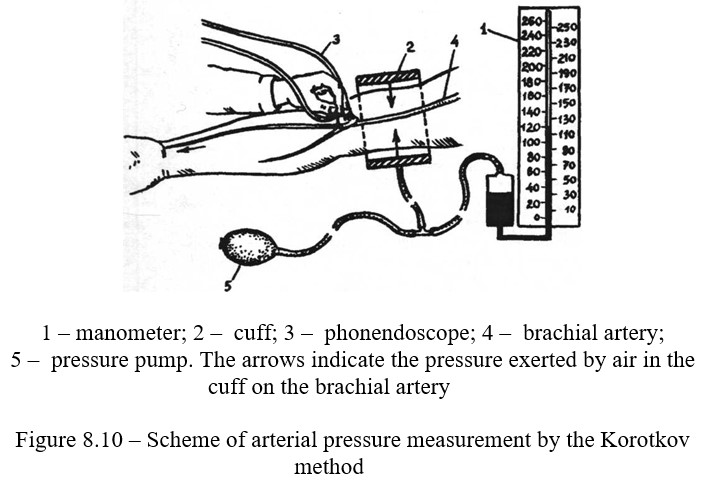
To determine the blood pressure in humans, various methods are suggested.
The purpose of the lesson: to get acquainted with the methods of bloodless determination of blood pressure in humans. To study changes in blood pressure during dosed physical work.
The following is necessary for work: testee, tonometer, phonendoscope, oscillograph
Work 1. Determination of blood pressure
Work progress: Rivva-Rocci method. Test person is placed sideways to the table, on which the hand is laid. Around the naked shoulder, a cuff is placed and strengthened, making sure that the shoulder is not squeezed tightly. On the palmar surface of the lower third of the forearm seek out a place of pulsation of the radial artery, followed during the study. Pump air into the cuff until the pressure in the manometer reaches 150-160 mm. Hg. In this case, the pulsation of the artery disappears.Slightly open the screw valve - the pressure in the cuff gradually decreases. Notice the readings of the manometer at the time of the pulse appearance, this value corresponds to the maximum blood pressure.
The method of Korotkov. Similarly, a cuff is applied. Then, in the area of the elbow fold of the testee, below the superimposed cuff, the membrane of the phonendoscope is placed above the brachial artery. No sound is heard. Air is forced into the cuff and pressurized (150-160 mm Hg), and then slightly opened with a screw valve, and the pressure in the cuff is gradually reduced.
At a certain pressure, a clear sound is suddenly heard. At this point it is necessary to look at the manometer reading. The pressure in the cuff, corresponding to the appearance of these sounds, is taken as maximum pressure (Figure 8.10). With further lowering of the pressure in the cuff, the audible sound becomes louder, and then disappears.
The manometer reading, corresponding to the moment of disappearance of sounds, is taken as minimum pressure. The value of the pulse pressure is calculated by subtracting the minimum value from the maximum pressure. The obtained values of the maximum, the minimum and pulse pressure should be recorded.
Work 2. Recording blood pressure in humans in an oscilloscope way
Work progress: the device of the oscilloscope allows to catch any movement of air in the sphygmomanometer, to strengthen it and to transfer it to the scribe.
Test person is placed sideways to the table, on which his hand is laid. Around the naked shoulder, the cuff of the oscilloscope is placed and strengthened. With the help of equalizing screws, set the ink-filled scribe so that it freely touches the paper of the cassette. Squeeze pressure pump injected air into the cuff until the pulsations of the radial artery cease.
Then open the screw valve and gradually lower the pressure in the cuff. The mercury level in the manometer will decrease, as a result, the cassette connected with it will gradually fall down and the scribe will record the corresponding waveform curve. On the oscillogram, the appearance of the first noticeable deflection corresponds to the maximum pressure. The maximum amplitude of the deflection will reach at the moment of average dynamic pressure of the artery.
When the pressure is minimal, the deflection of the oscilloscope drop sharply. Draw the received waveform. Find from the oscillogram the digital value of the maximum, minimum and average dynamic pressure.
Work 3. Indirect definition of venous pressure in the subcutaneous veins of the rear of the human hand
Work progress: the device for indirect determination of venous pressure consists of a capsule with a pelot on the rod, connected by means of a crane with a monolithic alcohol manometer. To measure pressure, select the bulging vein of the hand. The distal end of the vein is squeezed by the pelotis of the capsule, and the proximal end is squeezed by the finger with the blood squeezed to the side of the heart. Then slowly raise the capsule and, as soon as the vein begins to fill with blood, close the tap and on the scale of the manometer find the height of the venous pressure in mm Hg.
Control questions
- The value of blood pressure, its value in different parts of the vascular system. Factors on which the level of blood pressure depends.
- Physiological mechanisms of nervous regulation of blood pressure.
- Humoral mechanism of regulation of blood pressure. Mechanisms for regulating blood pressure when changing the position of the body, muscle work.
- Age changes in blood pressure. What factors determine the magnitude of the blood pressure?
- What methods are used to measure and record blood pressure in humans?
- Values and record of blood pressure.