9 Physiology of respiration
9.1 Spirometry and pneumography
Spirometry is a method of determining the vital capacity of the lungs. The vital capacity of the lungs (VCL) is the volume of air obtained by deep expiration after a preliminary deep inspiration.
The normal value of VCL is 2700-3000 cm3 for women, and 3500-4000 cm3 for men. The magnitude of the VCL varies depending on the height, age, degree of fitness, etc. For comparison of the actual VCL, i.e. VCL, as determined by given person, from the proper VCL, i.e. with VCL , which should be this person's normal, use the following simplified formula.
The required VCL is the number obtained from multiplying the growth (in centimeters) by 25 for men and by 20 for women. With normal breathing, the volume of inspired and expired air is much less than VCL, and on the average is 500 cm3. This is the so-called respiratory air. After a quiet expiration a person can always make a deep expiration. The air that can be expired after a normal expiration is called reserve air.
Its volume is on the average 1000-1500 cm3. After a normal inhalation, a person can in addition inspire 1500-1800 cm3 of air. This air is usually called the additional air. The sum of the volumes of the respiratory, reserve and additional air is VCL. In the lungs, even after the deepest expiration, there is always some air (1000-1500 cm3). This is residual air.
Thanks to the residual air, the lungs remain somewhat stretched even at the deepest expiration. The amount of volumes of VCL and residual air is called the lung volume. All the above volumes are called pulmonary volumes.
VCL and its components are determined by special instruments-spirometers and spirographs or by means of a primitive self-made spirometer.
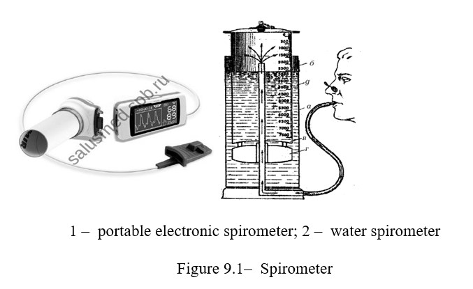
To determine VCL, water and air spirometers are used. Currently, portable spirometers are used with the ability to connect to a computer or printer. Such instruments allow performing respiratory tests, the results of which are directly read from the spirometer display, printed out with calculation of all the main parameters and indices (Figure 9.1).
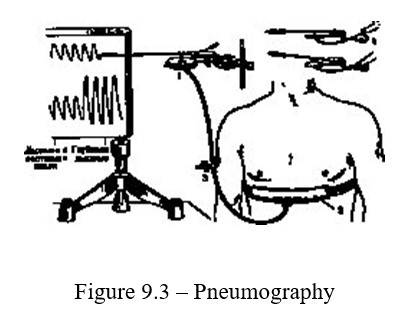
The recording of respiratory movements is recorded with the help of pneumograms of various systems. The principle of their action is that the respiratory movements are transmitted to the pneumograph and change the volume of the enclosed space available in it. These volume changes are recorded using the Marey capsule on the kymograph.
The purpose of the lesson: to get acquainted with the methods of spirometry, pneumography and human.
The following is necessary for work: spirometer, alcohol, cotton wool, calculator, pneumograph, kymograph, tripod.
Work 1 Spirometry
Work progress: VCL is determined by means of spirometer which consists of an outer and inner cylinder. The outer сylinder is filled with water to the mark "water level". The inner cylinder without a bottom, has a tire, is immersed in the inside of the outer one - into the water. In the center of the tire there is a hole covered with a rubber plug. To the wall of this cylinder is attached a scale graduated to 10,000 ml. Before the determination, the plug is opened and the inner cylinder is lowered until the zero digit of the scale coincides with the "reference level" mark. The mouthpiece is wiped with alcohol, and the hole is again closed with a plug. To determine the vital capacity of the lungs (VCL), the testee stands in front of the spirometer, straightens and, taking a deep breath from the atmosphere, takes a mouthpiece into his mouth, then makes the maximum expiration into the spirometer. The expiration is done slowly, without jerks. The volume of expired air is determined on a scale.
To determine the respiratory air, again the spirometer is brought to zero position, then after a quiet inspirationmake a quiet expiration in the spirometer. To determine the reserve air (RV=reserve volume) after a quiet inspirationmake a deep expire into the spirometer, which is in the zero position. Р
The value of the reserve volume of inspiration (RVinsp.) is determined by the next formula RVinsp = VCL - (AV+RVexp). Based on these three definitions, you can calculate the amount of additional air. To do this, it is necessary to combine the volumes of reserve and respiratory air and subtract this amount from the value of the vital capacity of the lungs.
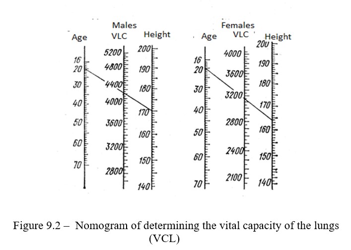
Determine the VCL, respiratory, reserve air volumes and calculate the volume of additional air. Determine the vital capacity of the lungs by the nanogram (Figure 9.2), knowing the age of the subject in years and his height in centimeters.
Work 2 Definition of the index of external respiration
Characterizes the potential of external respiration to remove
carbon dioxide and oxygen saturation of blood. It is determined by dividing ZHEL (ml) by body weight (kg). With prolonged absence of physical activity, the muscles participating in the respiratory movements lose some of their efficiency, which leads to a decrease in the effectiveness of gas exchange, a lack of oxygen in the body, and as a consequence, there is a risk of various pathologies.
The value, obtained by calculation, is compared with the tabular
(Table 9.1), evaluate the state.
Table 9.1 – The value of the respiratory rate in men and women
Estimation of state Definition of the index of external respiration value
|
|
Men under 40 years old |
Women under 40 years old |
|
Excellent |
66 and above |
62 and above |
|
Good |
61…65 |
58…61 |
|
Satisfactory |
56…60 |
51…57 |
|
The bad |
51…55 |
50…53 |
|
Very bad |
46…50 |
45…49 |
Calculate the main indicators of external respiration, record in the table
1 and compare with the proper values.
Work 3 Pneumography
Work progress: the rubber cuff is strengthened in front of the most moving part of the chest (middle third of the chest) (Figure 9.3). Slightly inflate the system with rubber pump through the tee-joint, making sure that too high pressure does not rupture the rubber membrane of the capsule.
Control questions
- The importance of breathing for the body. The main stages of breathing.
- The structure of the respiratory system.
- Ventilation of the lungs.
- External breathing. Methods of research. The mechanism of inspiration and expiration.
- Transport of respiratory gases by blood.
- Indicator of oxygen capacity of hemoglobin.
- Reflex regulation of respiration.
- Nervous-humoral regulation of breathing.
9.2 Gas exchange in the lungs and the role of dead space in breathing
Atmospheric air, passing through airways (larynx, trachea, bronchi, bronchioles) is cleared of dust, warmed and humidified. The air in the airways is not involved in gas exchange, so it is called the air of a "dead" or "harmful" space.
Only air that fills the pulmonary alveoless participates in gas exchange, which is in a state of continuous gas exchange with blood flowing through the capillaries of the lungs. Oxygen diffuses from the alveoles and blood, and carbon dioxide in the opposite direction. This is due to the difference in the partial pressures of the respective gases in the air on one side and in the venous blood on the other.
The partial pressure of oxygen in the alveolar air is 100-110 mm Hg. and in venous blood is 40.
The purpose of the lesson: to follow the diffusion of gases in the lungs and learn about the protective role of ciliated epithelium.
The following is necessary for work: Kipp's apparatus, a frog
Work 1 Diffusion of carbon dioxide in the lungs
Work progress: remove frog’s spinal cord and brain cord and attach it to the plate with their backs down. Cut the lower jaw. One of the edges of the slit is sewn with a thread and, pulling it by a string, insert a cannula into the trachea. Then, , a thread with a surgical needle is traced around the trachea and knotted, fixing the trachea on the cannula. The lungs through the canula slightly inflate and clamp a short rubber tube, clad on the free end of the cannula.
After that, open the thorax and gently separate the lungs from the body. The lungs are slightly inflated and placed in a bottle through which carbon dioxide is bubbled from Kipp's apparatus (Figure 9.4).
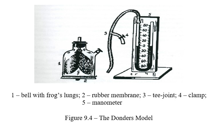
Now inside the lungs there is atmospheric air, outside - carbon dioxide. Gases diffuse through the wall of the lungs towards a smaller partial pressure, so nitrogen and oxygen from the lungs diffuse into the bottle, and carbon dioxide from the bottle - into the lungs. Carbon dioxide diffuses much faster than other gases, so the lungs gradually increase in volume, inflate and even burst.
Work 2 Experience with ciliated epithelium
The following is necessary for work: frog, dissection kit, plate, saline, pieces of finely chopped cork, slides, millimeter paper.
Work progress: the frog is immobilized in the usual way, the tongue is thrown upwards and exposed to the bottom of the oral cavity, whose mucosa forms folds and is covered with a ciliated epithelium.
The frog is fixed to the plate with the abdomen down. Then open the mouth widely and put a few pieces of cork on the folds of the mucosa. Be sure that they move towards the esophagus. Cut a piece of mucous membrane at the entrance to the esophagus (0.5 x 0.5 cm) and put epithelium down on the surface of the slide, moistened with physiological solution.Under the slide are placed millimeter paper and observe that a piece of mucous is moving in one direction. Measure the distance traveled by a piece of mucosa per minute.
Control questions
- Protective functions of the respiratory tract.
- Qualitative and quantitative indicators of respiration.
- Features of breath at the height.
- Features of breathing at depth.
- Methods of examining external respiration.
- Reflex self-regulation of respiration: the organization of the respiratory center and the role of its departments in the regulation of respiration. The autonomy of the respiratory center.
- The humoral mechanism of regulation of respiration (the role of carbonic acid, CO2, O2, H + ...).